
Download presentation, video, or reference.
Download presentation, video, or reference.
PARC ElectroCardioGram (Ultrasound the Heart) or PARCECG is a device for measuring electrical microalternations ECG. Microalternations ECG monitoring (primarily microalternations T-Wave ) - a relatively new non-invasive method of monitoring myocardial electrical instability caused little more than 20 years ago, entitled "Method MTWA" (Antonis A. Armoundas, Gordon F. Tomaselli and Hans D. Esperer Pathophysiological basis and clinical application of T-wave alternans / Journal of the American College of Cardioogy. - 2002; 40; P. 207-217.). Such devices are fundamentally different from standard ECG analyzers and interpreting ECGs, as measure and analyze not morphological features of ECG peaks (circuit analysis), and microscopic "trembling" of ECG signals. Microalternations calculated as low-amplitude vibrations of the ECG signal in successive contractions of the heart. Microalternations amplitude can be two orders of magnitude less than the standard ECG wave amplitude. Thus, the analysis of T-wave amplitude microalternations medium is 2...15mV, while the original wave amplitude T-200...700mV. In microalternations completely lost information about the features of the original amplitude of ECG waveform, i.e. alternans have the form of a random process, which does not contain the source of morphological features of ECG peaks in the analyzed lead.
Two Groups of Microalternations ECG
Class meters microalternations ECG can be divided into two groups. The first group includes gauges microalternations T-wave, the second - meters microalternations T-Wave + microalternations QRS-complex. The first group, in its development, came to a few realizations that instrument mainly are from foreign manufacturers. The second group is currently at the research stage and PARCECG is still the only instrument serial implementation of this technological direction. The reported groups have different clinical orientation (by appointment). Devices of the first group are mainly used to diagnose sick patients to assess the risk of malignant arrhythmias leading to sudden cardiac death. Among the most famous manufacturers of such devices can be called American firms Cambridge Heart ("SH2000") and GE ("GE Marquette"), as well as the German company MS Westfalia GmBh ("Case"). Meters T microalternations are either the main function of the device (CH), or option (GE). Measurement procedure in most cases associated with a load test and a long one. PARCECG due to a higher signal / noise ratio can measure microalternations even at rest in a very short time. Therefore, the main area of use - screening tool for early detection of progressive Amendments to the operational monitoring of the heart and myocardium.
The PARCECG Result
Chronologically PARCECG is one of the latest developments in the group microalternations meters, and different effective noise filtering system based on the "method of dispersion mapping." PARCECG designed for rapid surveys and requires only the signals of the limb leads with 4 electrodes recorded at rest for 30 seconds.
Meters microalternations ECG devices are providing new information on the most instrumental minor changes the electrical activity of the myocardium, which can not be detected by other methods. Average amplitude microalternations even physiologically normal myocardium are highly sensitive indicators of the cumulative effects of numerous physiological systems involved in the mechanisms of regulation of the heart. For example, PARCECG responds to subliminal metabolic changes that violate the synchrony of ventricular electrical excitation, microscopic changes in the ionic balance in myocytes, small shifts sympathetic-adrenal activation and other metabolic changes that are due to small quantities do not appear in the morphology of the ECG signals or ElectroCardioGram (ultrasound the heart). Similarly, PARCECG responds to the hidden dynamics of compensatory reaction of the left ventricle, which allows objective and timely manner to see an overload condition of the heart.
The main result of the high sensitivity of the instrument is that PARCECG ensures stable detection of adverse changes in the early stages of the disease, long before they are detected by other instrumental studies or manifest subjectively. PARCECG focused not on the type of pathology diagnosis and early detection of electrophysiological changes leading to a certain deviation from the norm or developing pathology.
The PARCECG System
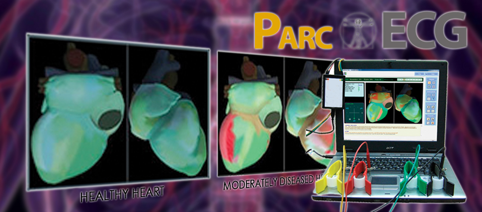
The PARCECG System is a new revolutionizing non-invasive diagnostic tool used for testing ischemic heart disease. It´s technology generates high quality 3D visuals projected from traditional ECG Dispersion Mapping. Dispersion Mapping is analyzing low amplitude oscillations of conventional ECG signals where we can generate a stable signal of ECG micro fluctuations by reflecting not just T-wave alterans (noise and artifact) but those of the QRS and R-wave complex as well. The 3-dimensional image projected by the system allows for physicians to observe the condition of the heart muscle and the intensity of ischemic heart disease.
General Method of Operation
- Four electrodes are applied in accordance to standard ECG arrangement of ECG limb leads
- ECG data acquisitioned in 30 seconds
- An image of the heart is formed on screen together with quantitative and qualitative analysis of cardiac electrical activity
The PARC ECG results are shown in the form of a numeric dispersive characteristics range and a dispersive mapping. The dispersive mapping is a colour-coded image of the heart in which green represented healthy and red represented pathological changes.
Dispersive characteristics are expressed by 9 analyzed groups of deviations. In these groups, the characteristics were analyzed reflecting electrophysiological abnormalities in the depolarization (generation of an electrical impulse that causes an action potential or a short lasting contraction of the heart muscles)and repolarization (heart muscle returns to its original state) of the myocardium.
The system therefore reacts to changes that exist or operate, below the threshold of normal ECG, that violate the synchrony of ventricular electrical excitation, as well as microscopic changes in the ionic balance of myocytes (muscle cell or muscle fiber) and other changes that are due to small quantities that do not appear in the morphology of the traditional ECG signals.
Career Opportunities to Health Practitioners, Therapists, & Medical Professionals
We offer a unique opportunity to effectively help operators be able to assist their customers and increase their revenue stream. This is an inexpensive device with very little recurring cost with a whole lot of added value. Currently signing up authorized agents, clinics, and doctors. This 30-SECOND test is changing the way people are diagnosed and treated. Contact us now for information and registration.
Be the First to Get this Ground-Breaking Technology!
Our Package is a Turnkey System. We offer training and support through videos and power point presentation as well as online training and support via webinars and using other support tools like teamviewer for immediate help. This device can also be used in Hospitals and many companies are now asking us to private label this device to suit their needs including those like franchised wellness centers and network marketing companies in the health care industry. If you are looking to increase your revenue stream this might just be the device for you or your company.
Why say ´YES´ to PARCECG?
Feature: It uses ECG dispersion mapping by capturing low amplitude waveforms ,to project a 3-dimensional image of the heart , and highlights impaired areas of the myocardium.
Benefit: This allows physicians to assess their patients in real time for ischemic heart disease. This shortened time allows for faster diagnosis by our doctors and thereby allowing for faster medical administration to their patients.
Feature: Uses ECG wave alterans (noise) for information.
Benefit: This specific feature is what this system monopolizes on. It´s ability to use information that is regarded as ´insignificant or unusable´ by the traditional ECG systems, is what sets it apart from the rest.
Feature: Uses four limb lead wires with patients sitting in an upright position and fully clothed.
Benefit: This particular feature of the Parc ECG allows for patients being more comfortable during the procedure. Many may prefer this alternative rather than being in a state of undress.
Feature: Specificity and sensitivity to ischemic heart disease increased by 80%.
Benefit: Improved sensitivity to detect early warning signs will thereby allow for detection of extremely small deviations during cardiac cycles. This is important since these small deviations are generally invisible to regular ECG systems.
The characteristics of PARC ECG amounts to greater possibilities than traditional analogues used by medical practitioners today. The very idea of seeing a 3-dimensional projection of all the raw data gives it the unique and phenomenal status that it deserves, that is, one that surpasses the expectations of a medical practitioner , and in the interim, accomplishes its job of providing quality patient care and services. PARCECG, paving the way for new age diagnostic technology that changing western medicine as we know it.